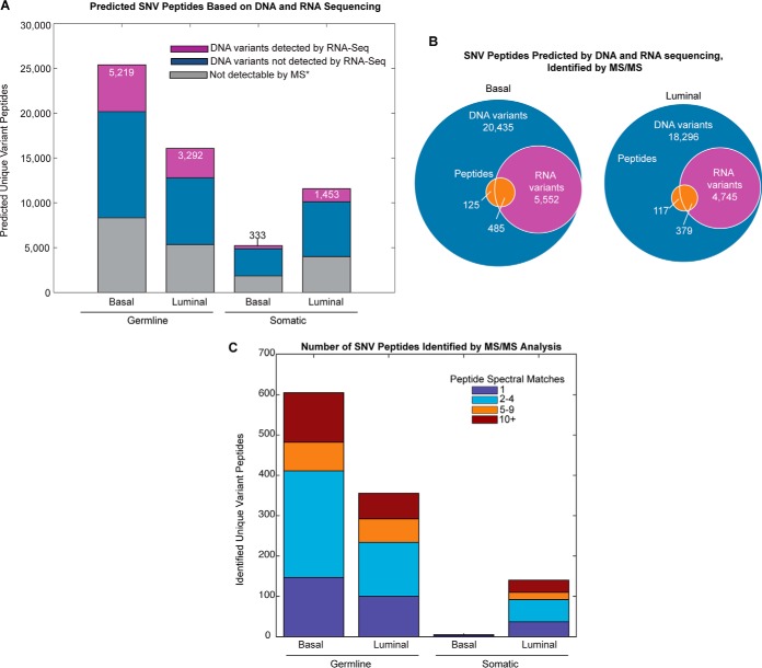
Fig. 2.
Single nucleotide variant (SNV) peptide expression in basal and luminal tumors. (A) Predicted proteomic change based on single nucleotide variants. Purple bars show the number of predicted unique peptides based on DNA variants that were also detected by RNA-Seq, blue bars indicate the number of predicted unique peptides based on DNA variants that were not detected by RNA-Seq, and gray bars indicate those peptides that are not detectable by mass spectrometry techniques due to size limitations (*peptides that have lengths <6 or >30 amino acids). (B) Proportion of DNA variants that were also identified by RNA sequencing and MS/MS proteomics for luminal and basal breast tumors. (C) Total basal and luminal variant peptides identified by MS/MS. Identification by iTRAQ, label-free MS/MS, or both is indicated by stacked bar color.

