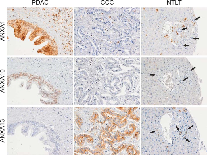
Fig. 3.
Representative immunohistochemical staining of PDAC, CCC, and nontumorous liver tissue (NTLT) with antibodies against the annexins A1, A10, and A13. ANXA1 presented with a strong nuclear and diffuse cytoplasmic staining of PDAC cells whereas most tumorous and all nontumorous cholangiocytes (indicated by arrows) were unstained. In addition, stromal and inflammatory cells often expressed high amounts of ANXA1. In the case of ANXA10, a nuclear staining of PDAC cells varying in intensity was observed. CCC cells showed none or weak staining and nonmalignant cholangiocytes were completely unstained. ANXA13 was more often expressed in CCC than PDAC cells as well as in nontumorous bile duct cells presenting low to moderate expression, especially in the luminal membranes but also often in the cytoplasm. Hepatocytes did generally not express ANXA1, ANXA10, or ANXA13. Original magnification 400x.

