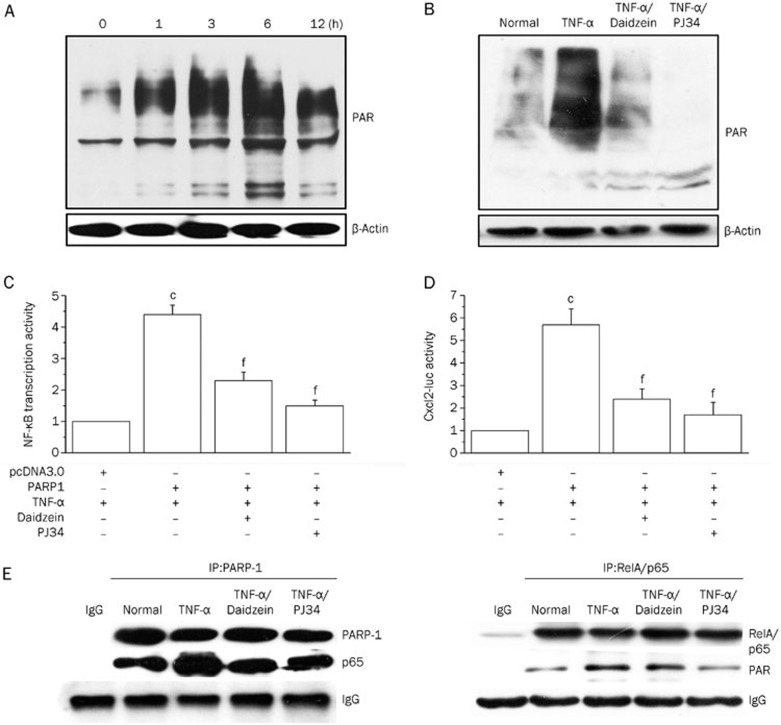
Figure 3.
Daidzein blocks pro-inflammatory gene expression via inhibiting PARP-1 activity. (A) MLE-12 cells were normally cultured or stimulated with TNF-α for different times, as indicated, or (B) the cells were normally cultured or stimulated with TNF-α (in the presence or absence of PJ34 or daidzein) for 6 h. The cells were lysed, the lysates (20 μg protein) were resolved using SDS-PAGE, and protein PARylation was detected using the anti-PAR antibody. (C and D) MLE-12 cells were co-transfected with the PARP-1 expression plasmid (or the negative control pcDNA3) with the reporter plasmids NF-κB-luc or Cxcl2-luc, as indicated. After stimulation with TNF-α with or without inhibitors, a dual reporter assay was performed as described above. The transcriptional activity of NF-κB or Cxcl2 in cells transfected with the control plasmid (pcDNA3) was considered the baseline and valued as 1. (E) Cells (1×10⁷) were stimulated with TNF-α in the presence or absence of daidzein or PJ34 for 6 h, and immunoprecipitation was performed. Antibodies against PARP-1 (left panel) or RelA/p65 (right panel) were used to obtain complexes that were resolved using SDS-PAGE. Immunoblotting was performed using antibodies specific for RelA/p65, PARP-1 and PAR, as indicated. Data (mean±SD) are representative of three independent experiments (cP<0.01 compared with cells transfected with pcDNA3; fP<0.01 compared with TNF-α-stimulated cells).

