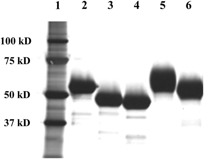
Fig. 2.
SDS–PAGE of the purified rhATs. The purified rhATs were reduced with dithiothreitol and 2 µg of each sample was run in the gels. Electrophoresis was performed in the presence of sodium dodecyl sulfate, and the samples were detected by silver staining. Protein standards (lane 1), phAT (lane 2), rhAT-Manα (lane 3), rhAT-Manβ (lane 4), rhAT-Comα (lane 5) and rhAT-Comβ (lane 6) were analyzed.

