Figure 1.
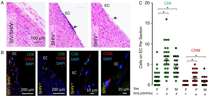
Simian immunodeficiency virus (SIV) or simian-human immunodeficiency virus (SHIV) infection in rhesus macaques (RMs) is associated with focal endothelial proliferation and subendothelial migration of inflammatory cells. Sections of formaldehyde-fixed paraffin-embedded descending thoracic aorta from uninfected and SIV/SHIV-infected were processed for hematoxylin-eosin (HE) histochemical, and immunofluorescence staining using anti-CD68, anti-CD8, and DAPI. A, B, Representative data obtained by microscopic examination (×20, ×40, and ×100 objectives) of aortic endothelium from 16 infected and 16 uninfected animals are shown, indicating endothelial adhesion/infiltration of immune cells (arrow) in the aorta of an infected animal (A; HE staining). B, CD8+ and CD68+ cells identified by immunofluorescence staining were detectable more frequently on or below the endothelium of SIV/SHIV-infected animals. C, Summary data were generated by counting the numbers of CD8+ (green) and CD68+ (red) cells adhered to the aortic endothelium of SIV-infected (open circles), SHIV-infected (black circles), and uninfected male (M) and female (F) animals. Five aortic sections were examined from each of 16 infected and 16 uninfected control animals. *P < .001; 2-tailed nonparametric Mann–Whitney U test. Abbreviations: DAPI, 4',6-diamidino-2-phenylindole; EC, endothelial cell.

