Fig. 3.
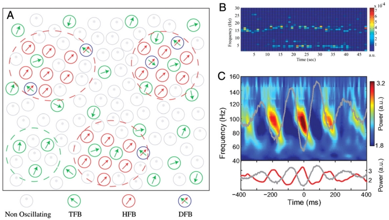
Multiplexing and cross-frequency interaction. [A] Functional organisation of a model of the STN in Parkinson’s disease depicting neurons classified by their oscillatory activity. Neurons are described as oscillating at tremor frequency band (TFB), high-frequency [8–20 Hz] band (HFB) or dual-frequency band (DFB). The model involves multiplexing both at the nuclear and neuronal levels. [B] Example of the time–frequency firing pattern of a DFB neuron oscillating simultaneously at both tremor and high (beta-range) frequencies; clear evidence of multiplexing within a neuron. [C] Cross-frequency coupling between theta-phase and gamma-amplitude in the rat striatum obtained during navigation of a T-maze task.
[A] and [B] adapted with permission, Moran et al. (2008), [C] adapted with permission, Tort et al. (2008).

