Figure 1.
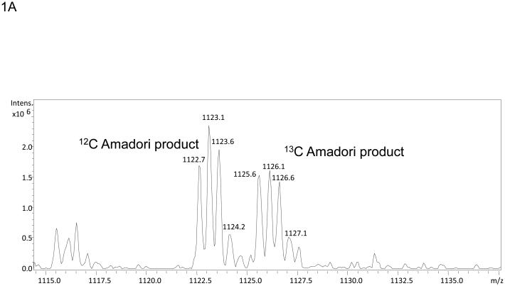
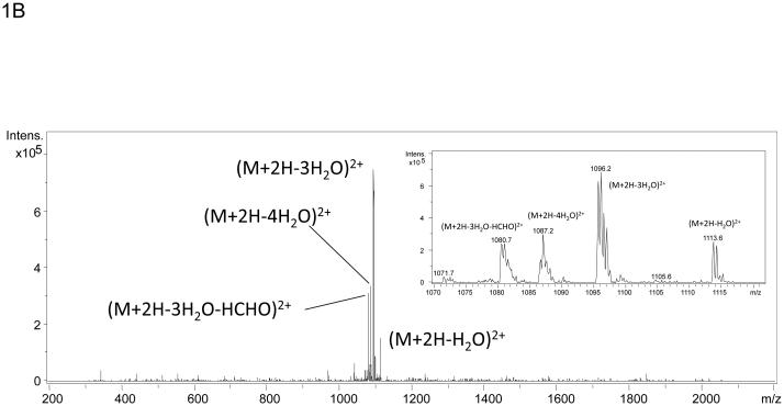
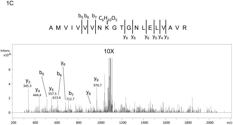
Mass spectra of the tryptic peptide spanning amino acids 452-471from glycated recombinant Ara h 1. Panel A shows the mass spectrum of the dyad of ions at m/z 1122.7 and m/z 1125.6 that correspond to amino acid residues 452-471 with K460 modified by glycation that arises from the incubation of rAra h 1 with 1:1 12C glucose:13C glucose. Panel B is the low information MS/MS spectrum of ion m/z 1122.7 with an inset showing a zoom in from m/z 1070 to m/z 1120. The neutral losses of 3 waters and 3 waters plus HCHO are characteristic for Amadori product modified peptides. Panel C is the 10X zoom of the MS/MS of ion m/z 1122.7 which shows very low abundance b- and y- series ions.


