Figure 1.
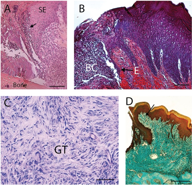
Cellular and histologic events involved in gingival wound healing. (A) Tissue section shows the inflammatory phase of gingival wound healing. Arrow indicates inflammatory cells. SE, sulcular epithelium. Tissue section was stained with eosin-hematoxylin. Bar = 250 µm. (B) Micrograph shows the migration of epithelial cells (E) during wound healing. BC, blood clot. Tissue section was stained with phosphotungstic acid hematoxylin. Bar = 250 µm. (C) A more advanced stage of wound healing shows the formation of granulation tissue (GT). Tissue staining was stained with Giemsa. Bar = 50 µm. (D) Section represents the tissue-remodeling phase. Wound has been replaced with connective tissue (arrow). This tissue has a high proportion of cells. The gingival epithelium has covered the wound. Section was stained with mallory trichrome. Bar = 250 µm.

