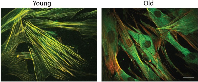
Figure 2.
Aging affects myofibroblastic differentiation. These images show human gingival myofibroblasts derived from young and aged donors stimulated with transforming growth factor–β1 (TGF-β1) for 72 h. In both cases, young and aged cells expressed the actin isoform α–smooth muscle actin (α-SMA) (green staining). However, α-SMA was incorporated into actin stress fibers (red) only in fibroblasts derived from young individuals. In aged cells, α-SMA (green) remained as a cytosolic staining, as shown in this image. These results suggest that aged myofibroblasts were not able to form mature actin fibers. These defects might affect tissue remodeling during wound healing. Bar = 10 µm. For details, see Cáceres et al. (2014). This figure is available in color online at http://jdr.sagepub.com.

