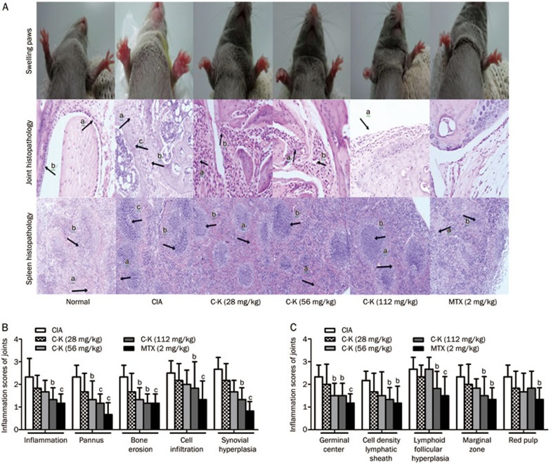
Figure 3.
Effects of C-K on the histopathology of the spleen and joints of CIA mice. A photomicrograph of joint histopathology showing synoviocyte hyperplasia (arrow a), blood vessels (arrow b), articular cartilage destruction and pannus (arrow c). A photomicrograph of spleen histopathology showing red pulp congestion (arrow a), white pulp proliferation (arrow b) and germinal center formation (arrow c) (A). Effects of C-K on joint histopathology (B). Effects of C-K on spleen histopathology (C). Mean±SD. n=10. bP<0.05, cP<0.01 vs CIA mice.

