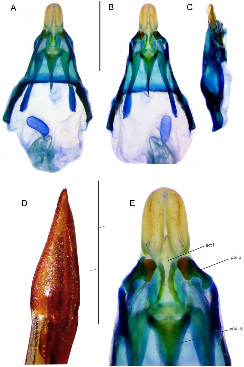
Fig 4. Genitalia of Pixibinthus sonicus.
(A) Dorsal, (B) ventral, and (C) lateral views of male genitalia. (D) Lateral view of apex of female ovipositor. (E) Ventral view of male genitalia. For abbreviations and symbols, see Material and methods. Scale bars = 1 mm.

