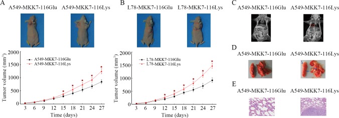
Fig 3. The effects of MKK7 p.Glu116Lys on tumor growth and metastasis.
(A, B). Subcutaneously implanted MKK7-116Glu (A549-MKK7-116Glu and L78-MKK7-116Glu cells) and MKK7-116Lys (A549-MKK7-116Lys and L78-MKK7-116Lys cells) cells xenografted tumors were established and were observed for a total of 4 weeks. Tumor volumes represented the mean ± SD of 6 mice per group. Columns, mean; bars, SD. The symbol “★” indicated a statistical significance with P < 0.05 between the cells transfected with the two different transfectants. (C-E). A549-MKK7-116Glu or A549-MKK7-116Lys cells were separately injected into the tail vein of each mouse. After proximately 10 weeks, lung metastases were evaluated using magnetic resonance imaging, macroscopic observation and histomorphology under microscopy. The red loops and arrows indicate the metastases.

