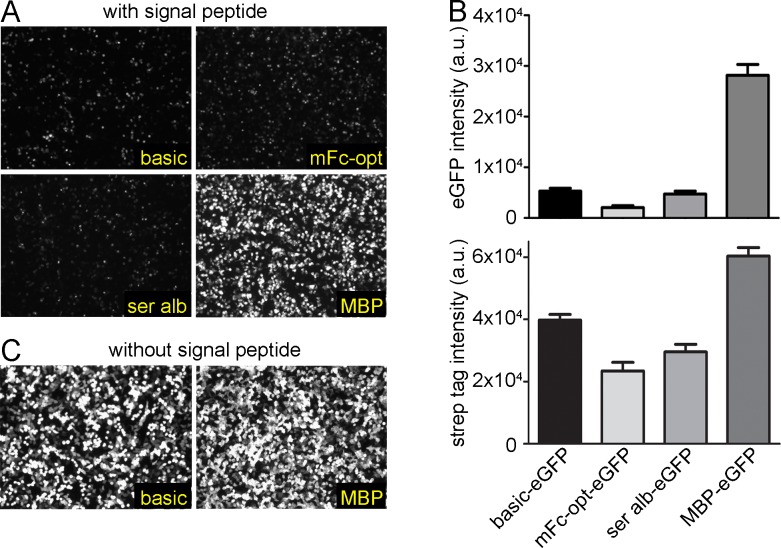
Fig 3. Enhanced expression of eGFP.
HEK293 cells were transiently transfected with eGFP alone (basic) or fused to different expression tags. (A) All proteins contained a signal peptide sequence for secretion. The eGFP signals were visualized by fluorescence microscopy. (B) To quantify the secreted eGFP, the supernatants from the HEK293 cells were excited with a laser at 488 nm and the emission signal was detected at 509 nm (top). Densitometric analysis of secreted eGFP from HEK293 cells by western blot analysis using a Strep-Tactin®-HRP conjugate to detect Strep II tagged eGFP fusion proteins (bottom). (C) Without signal peptide, eGFP was transiently expressed intracellularly with or without fused MBP. eGFP signals were visualized by fluorescence microscopy. (a.u.: arbitrary unit).

