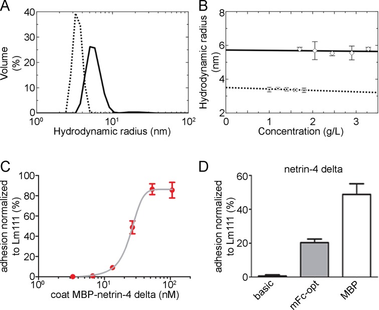
Fig 5. Biophysical analysis of MBP as well as MBP fusion proteins and cell attachment studies.
(A) Light scattering profile obtained for a 1.80 mg/mL solution of MBP (dotted line) and for a 3.28 mg/mL solution of MBP-netrin-4 delta (solid line) in the TBS buffer display the respective hydrodynamic radius. (B) Concentration dependence of the hydrodynamic radius of MBP (dotted line) and MBP-netrin-4 delta (solid line), respectively, deduced from the peaks of DLS profiles. (C-D) B16-F1 cells were allowed to adhere to surface coated with MBP-netrin-4 delta. (C) Dependence of cell attachment on the coating concentration of MBP-netrin-4 delta. (D) Cell attachment at E50 coating concentration (MBP-netrin-4 delta) for different netrin-4 delta proteins (with and without tag).

