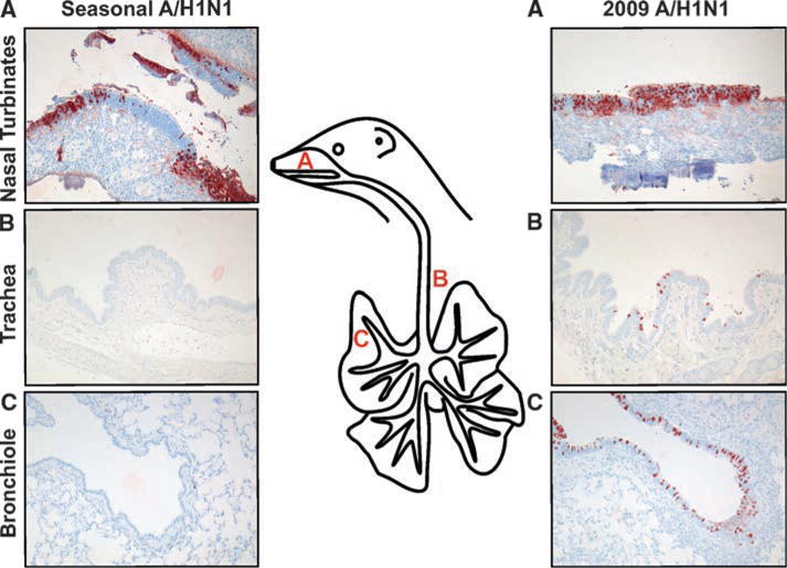
Fig. 2.

Immunohistochemical analysis of respiratory tract tissues of ferrets inoculated with seasonal or 2009 A(H1N1) influenza virus, collected at 3 days after inoculation. Tissue sections of the nasal turbinates (A), trachea (B), and bronchi (C) were stained with a monoclonal antibody against influenza A virus nucleoprotein, which is visible as a red-brown staining. In animals inoculated with seasonal influenza virus, only cells in the nasal turbinates stained positive for nucleoprotein, whereas in animals inoculated with 2009 A(H1N1) influenza virus, cells in the nasal turbinates, trachea, and bronchi stained positive. See fig. S2 for data taken at 7 days after inoculation.
