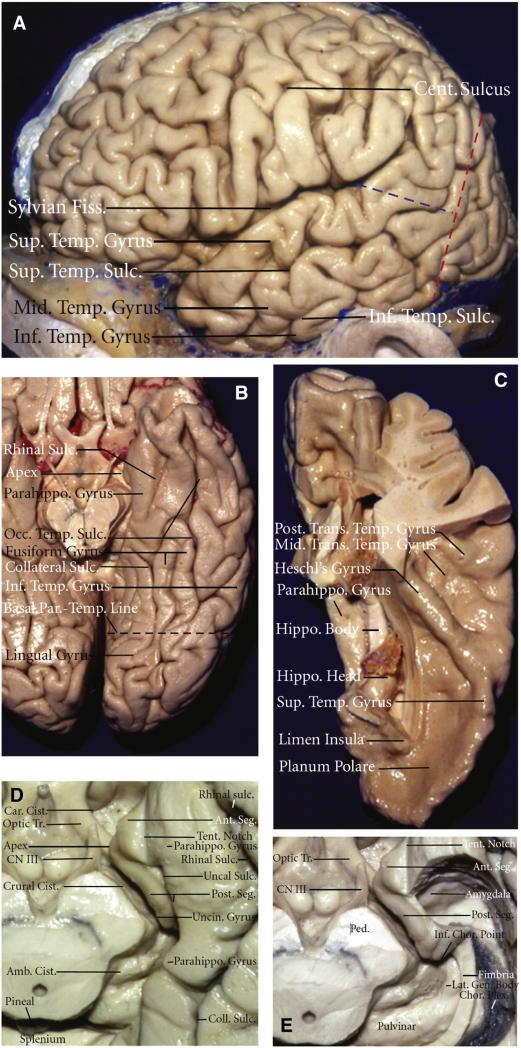
Fig. 1.
Microsurgical anatomy of the temporal lobe. A) Lateral view of the left hemisphere. The lateral surface of the temporal lobe consists of three parallel gyri: superior, middle, and inferior temporal gyri. These gyri are separated by the superior and inferior temporal sulci. The lateral parietotemporal line (red dashed line), an imaginary line connecting the preoccipital notch and parietooccipital sulcus, separates the temporal and occipital lobes, and the occipitotemporal line (blue dashed line), an imaginary line connecting the posterior margin of the sylvian fissure with lateral parietotemporal line, separates the temporal and parietal lobes. B) Inferior view of the left temporal lobe. The basal surface of the temporal lobe consists of, from lateral to medial, the inferior margin of the inferior temporal gyrus, the fusiform gyrus, and the parahippocampal gyrus. The fusiform gyrus is separated laterally from the inferior temporal gyrus by the occipitotemporal sulcus and medially from the parahippocampal gyrus by the collateral posteriorly and rhinal sulci anteriorly, which are not continuous in every case. The basal parietotemporal line connecting the preoccipital notch and inferior end of parietooccipital sulcus separates the temporal and occipital lobes at the basal surface. C) The superior view of the left temporal lobe. This surface facing the sylvian fissure is divided, from anterior to posterior, into three portions: the planum polare, the anterior transverse temporal gyrus, referred to as the Heschl's gyrus, and the planum temporale containing the middle and posterior transverse temporal gyri. D) Enlarged view of the anterior and middle segments of the medial temporal region. The anterior segment of the uncus faces the carotid cistern, and the posterior segment faces the crural cistern and the cerebral peduncle. The uncal apex is positioned lateral to the oculomotor nerve. The cortical component of the middle medial temporal region formed by the parahippocampal gyrus faces the midbrain across the ambient cistern. E) The medial temporal region with hippocampus and dentate gyrus having been removed while preserving the fimbria and the choroid plexus attached along the choroidal fissure. The amygdala forms the anterior wall of the temporal horn and fills most of the anterior segment of the uncus. The inferior choroidal point, located at the lower end of the attachment of the choroid plexus in the temporal horn, is positioned behind the head of the hippocampus, anterior to the lateral geniculate body, and lateral to the posterior edge of the cerebral peduncle. Amb.: ambient; Ant.: anterior; Calc.: calcarine; Car.: carotis; Cent., central; Chor.: choroidal; Cist: cistern; Coll.: collateral; CN III: oculomotor nerve; Fiss.: fissure; Gen.: geniculate; Hippo.: hippocampus; Inf.: inferior; Lat.: lateral; Mid.: middle; Occ.: occipital; Parahippo.: parahippocampal; Par.-Occip.: parietooccipital; Par.-Temp.: parietotemporal; Ped.: cerebral peduncle; Post.: posterior; Seg.: segment; Sulc.: sulcus; Sup.: superior; Temp.: temporal; Tent.: tentorial; Tr.: tract; Trans.: transverse; Uncin.: uncinate. (For interpretation of the references to color in this figure legend, the reader is referred to the web version of this article.)
Figure and legend modified and reproduced with permission from Kucukyuruk et al. [24] distributed under the Creative Commons Attribution License.

