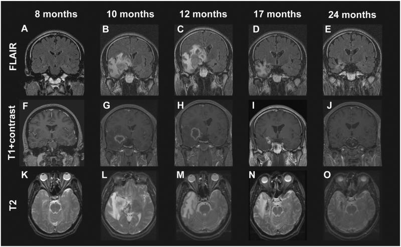
Fig. 5.
Development of radiologic changes in a patient with mesial temporal lobe epilepsy treated with a 24-Gy dose gamma knife stereotactic radiosurgery. FLAIR (A–E) and T2 (K–O) hyperintensity appeared within the medial temporal lobe beginning by the 10th postoperative month and peaked in intensity at 12 months, corresponding to a decline in the proportion of patients experiencing complex partial seizures. Contrast enhancement (F–J) followed a similar time course, except that it preceded T2 changes and diminished quickly after months 10–12. Enhancement was typically ring-enhancing and centered over the target region.
Figure and legend reproduced with license and permission from Chang et al. [73].

