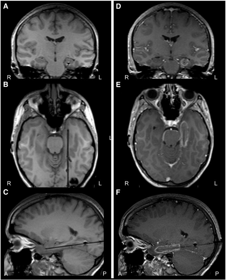
Fig. 6.
Stereotactic laser thermo-ablation in mesial temporal lobe epilepsy. A–C) T1-weighted periprocedural MRI coronal (A), axial (B), and sagittal (C) images showing laser probe placement along the axis of the left hippocampus, prior to thermo-ablation in a patient with mesial temporal lobe epilepsy. D–F) Contrast-enhanced T1-weighted MRI coronal (D), axial (E), and sagittal (F) images after thermo-ablation of mesial temporal lobe structures, with contrast enhancement observed in the region of ablation. A: anterior; L: left; P: posterior; R: right.

