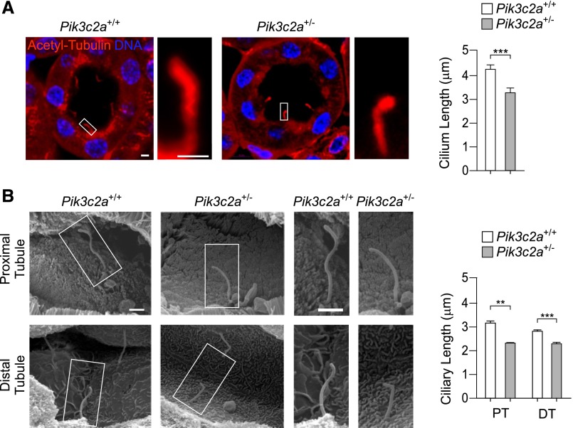
Figure 4.
Pik3c2a+/− mice show shorter cilia in proximal and distal renal tubules. (A) Representative micrographs of cilia in wild-type and Pik3c2a+/− kidney tubules, detected by immunostaining with acetylated-α-tubulin (red) and DNA (DAPI, blue). Bar=2 μm. Graph shows the quantification of ciliary length in tubules of wild-type and Pik3c2a+/− mice. n=100 cilia per /genotype. (B) Imaging by scanning electron microscopy of proximal and distal tubule primary cilia in kidneys of 6-week-old wild-type and Pik3c2a+/− mice. Quantification of 20 (proximal tubule, PT) and 75 (distal tubule, DT) cilia per genotype are provided on the right. Bar=1 μm.

