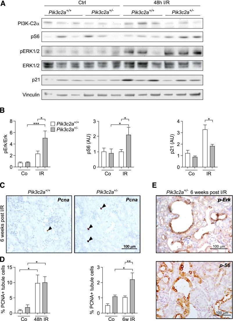
Figure 6.
Uncontrolled proliferative signaling in the tubules of Pik3c2a+/− kidneys after I/R. (A and B) Western blot analysis of pathways regulating proliferation in wild-type and Pik3c2a+/− samples from either control (ctrl) or ischemic (I/R) kidneys at 48 hours after treatment. Activation of the MAPK pathway is investigated through analysis of pErk, while phosphorylation of S6rp shows the activation of the mTOR pathway. p21 is a cyclin-dependent kinase inhibitor upregulated after ischemic injury of the kidney. A representative western blot (A) as well as protein quantification (B) of n=8 wild-type and n=8 Pik3c2a+/− mice are shown. (C and D) Analysis of proliferation was achieved by immunohistochemical staining of PCNA in sections of either control (ctrl) or ischemic (I/R) kidney from wild-type and Pik3c2a+/− mice. Representative sections of a wild-type and a Pik3c2a+/− kidney at 6 weeks after I/R is shown (C). Graphs provided in (D) represent the percentage of tubular cells showing a PCNA positive nucleus over the number of tubular cells per section at either 48 hours or 6 weeks after I/R. For all groups n=6 mice. (E) Representative micrographs showing that the majority of cystic lesions is positive for pErk and pS6rp in sections from a Pik3c2a+/− kidney at 6 weeks after I/R. PCNA, proliferative cell nuclear antigen.

