Figure 8.
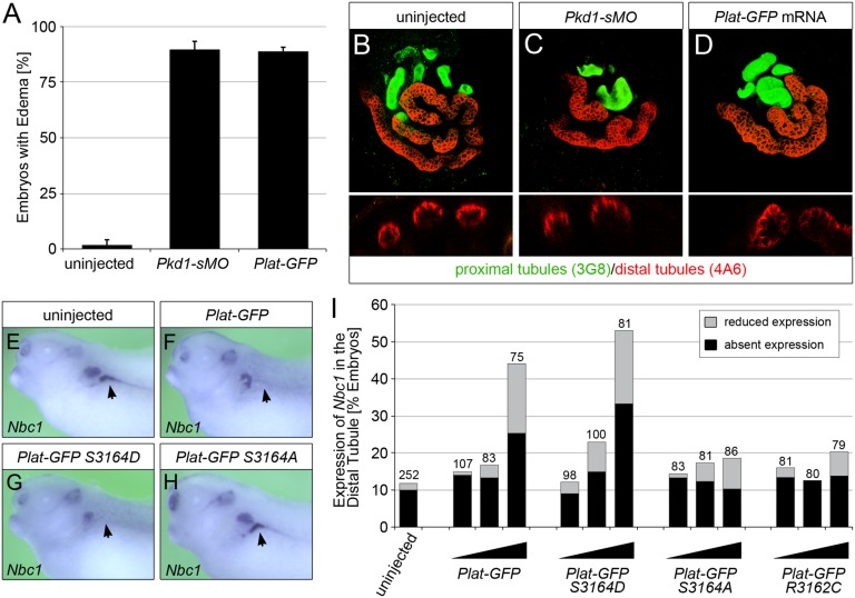
Ectopic expression of PLAT-GFP causes PKD in Xenopus embryos. (A) Xenopus embryos injected radially with a total of 3.2 pmol Pkd1-sMO or 8 ng Plat-GFP mRNA as well as uninjected control embryos were analyzed at stage 42 by morphology. Bar diagram shows the percentage of embryos with edema as a sign of kidney dysfunction. Error bar corresponds to SD of multiple experiments. (B–D) Whole–mount immunofluorescence analysis with 3G8 (green) and 4A6 (red) of the embryos in A to visualize proximal tubules as well as distal tubules and duct, respectively. Upper panels show three-dimensional reconstruction of z stacks, whereas lower panels are cross-sections through individual proximal tubules. (E–I) Xenopus embryos were injected at the four-cell stage into a single blastomere with increasing amounts (0.5, 1.0, and 2.0 ng) of wild-type Plat-GFP, Plat-GFP(S3164D), Plat-GFP(S3164A), and Plat-GFP(R3162C). Expression of Nbc1 in the DT1 distal tubular segment was examined by whole–mount in situ hybridization comparing the injected and the contralateral noninjected pronephri at stage 39. E–H show representative embryos (Nbc1 staining in the DT1 segment is indicated by arrows). (I) Quantification of three independent experiments depicting the percentages of embryos with absent and reduced Nbc1 expressions. The total number of embryos analyzed is indicated above the bars.

