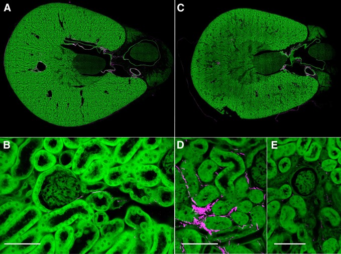
Figure 18.
Second harmonic generation sections showed similar patterns to Sirius red with possible mild increases in interstitial collagen in CP mice. (A and B) Control mouse MPM/SHG of kidney showing minimal collagen (magenta), almost exclusively in perivascular orientation. (C) Low-power view CP-treated kidney does not show marked differences compared with control. (D) High power view shows some interstitial peritubular collagen (magenta), but the collagen was not associated with abnormal glomeruli or "atubular" glomeruli as in part E. Scale bar=50 μm.

