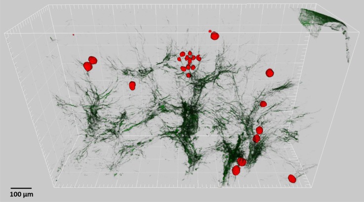
Figure 20.
The collagen fibrosis in CP kidneys is not co-localized with atubular glomeruli. Atubular glomeruli from this CP mouse kidney are pseudocolored in red. Second harmonic collagen signal is dark green. The atubular glomeruli are distributed throughout the kidney section and span cortex to medulla. A small cluster of small atubular glomeruli is evident in center.

