Figure 1.
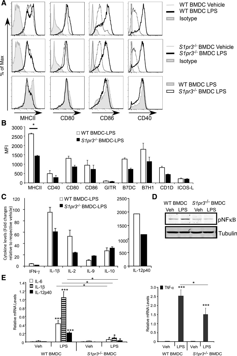
LPS–treated S1pr3−/− BMDCs produce fewer cytokines and costimulatory molecules compared with WT BMDCs. WT and S1pr3−/− BMDCs were treated with vehicle (PBS) or 100 ng/ml LPS for approximately 20 hours. (A) Flow cytometry histograms (overlays) illustrating percentage of maximum signal for MHCII, CD80, CD86, CD40, or control isotype IgG when gating on CD11c+ cells. (B) MFI calculated from FACS histograms. (C) Changes in protein expression levels of cytokines (mouse 30–plex Luminex) in supernatants collected from WT and S1pr3−/− BMDC cultures after incubation with PBS or LPS for 20 hours. (D) Western blot of phosphorylated NFκB (pNFκB) in PBS– (vehicle [Veh]) or LPS–treated WT or S1pr3−/− BMDCs. (E) Changes in mRNA expression of proinflammatory cytokines from WT and S1pr3−/− BMDCs treated with Veh (PBS) or 100 ng/ml LPS for 3 hours (values expressed relative to GAPDH). Values are mean±SEM (n=2–3 mice with three replicates for each experimental set). *P<0.05; ***P<0.001.

