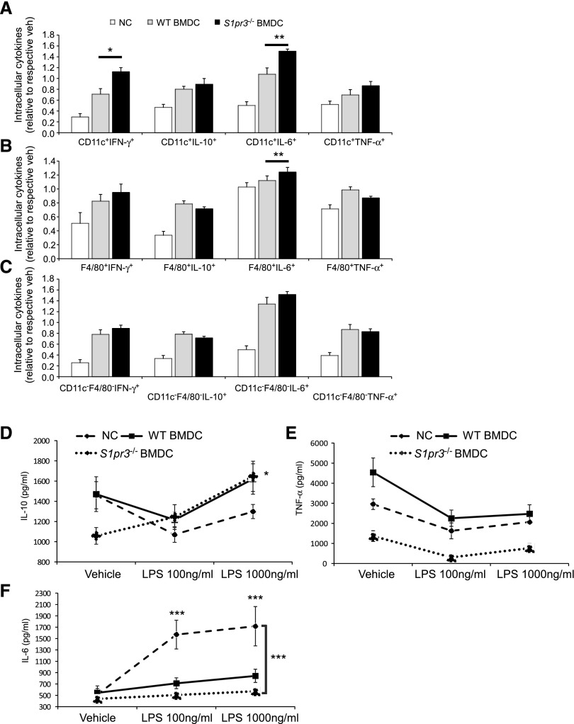
Figure 4.
Splenocytes from S1pr3−/− BMDCs treated mice produce less inflammatory cytokines. Characterization of splenocytes from mice injected with no cells (NCs), WT BMDCs, or S1pr3−/− BMDCs. Splenocytes were isolated 24 hours after BMDC injection; 1×10⁵ splenocytes per well were treated with 100 or 1000 ng/ml LPS for an additional 24 hours. (A–C) Splenocytes were restimulated with PMA (10 ng/ml), ionomycin (2 μg/ml), and brefeldin A (5 μg/ml) for an additional 5 hours; intracellular cytokines (IFN-γ, IL-10, IL-6, and TNF-α) were measured by flow cytometry in (A) CD11c+ cells, (B) F4/80+ cells, and (C) CD11c−F4/80− (double negative) cells. Values are mean±SEM (n=3). *P<0.05; **P<0.01. (D–F) Secreted levels of cytokines were measured by ELISA at 24 hours after vehicle or LPS (100 or 1000 ng/ml) in supernatants. (D) IL-10 (values are mean±SEM; n=3). *P<0.05, S1pr3−/− BMDC (vehicle compared with 1000 ng/ml LPS). (E) TNF-α and (F) IL-6 (values are mean±SEM; n=3). ***P<0.001, NCs (vehicle compared with both LPS doses); S1pr3−/− BMDC–treated splenocytes with LPS compared with NC LPS–treated splenocytes at both LPS doses.

