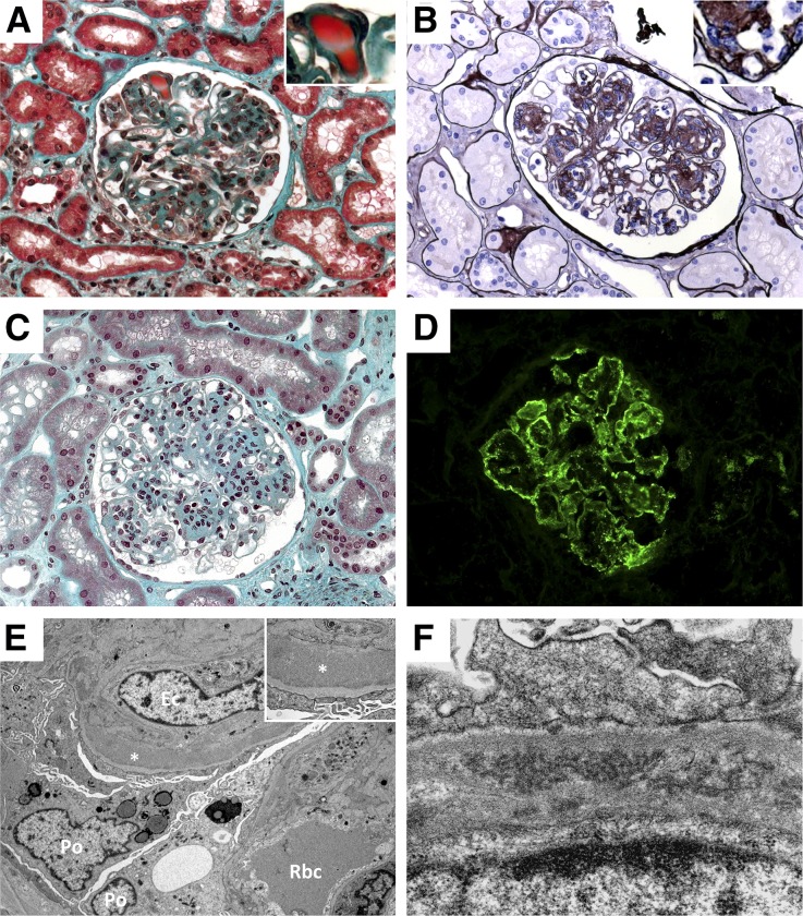
Figure 1.
Glomerular lesions in the course of noninfectious MCV. Light microscopy examination showing typical MPGN with endocapillary proliferation, capillary lumen infiltration by monocytes–macrophages, and Ig-related protein thrombi in the capillary lumen (A, Masson’s trichrome stain, ×400), typical doubles contours (B, Jones’ staining, ×400) and a case of mesangial proliferative GN (C, Masson’s trichrome stain, ×400). Immunofluorescence study showing granular and immune-complex glomerular deposits composed of IgG (D). IgM, and both κ and λ light chains were also detected in a similar pattern (data not shown). Electron microscopy analysis showing capillary thickening and subendothelial granular dense deposits (E; Po, podocyte; Ec, endothelial cell; Rbc, red blood cell; *, dense deposits), and organized microtubular deposits (F).

