Abstract
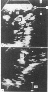
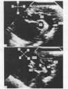
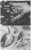
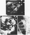
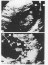
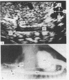
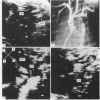
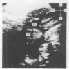
To establish an integrated non-invasive method for diagnosing coarctation, cross-sectional echocardiographic appearances of 48 neonates and infants with coarctation were combined with clinical information on the peripheral pulses. Measurements of the ascending aorta, aortic arch, and isthmus were made and compared with those from controls matched for weight and age. Confirmation of the coarctation was available in all cases. Angiocardiographic measurements were performed in 15 patients from either the group with coarctation or the controls. After the aortic arch had been analysed segment by segment 40 patients were found to have preductal coarctation, five juxtaductal coarctation, and three postductal coarctation. In one of the patients in the latter group the obstruction was situated in the abdominal aorta. Specific echocardiographic features were present in each subgroup. Echocardiographic measurements were about two thirds of those obtained by angiocardiography. By combining information on the peripheral pulses, isthmic size, and the presence of a discrete shelf in the aorta it was retrospectively possible to predict correctly the presence of coarctation in 45 out of 48 cases. Since the beginning of this study 29 patients have undergone surgery without prior invasive investigation. A combination of clinical assessment and cross-sectional echocardiographic features allows a reliable diagnosis of coarctation to be made in most cases.
Full text
PDF
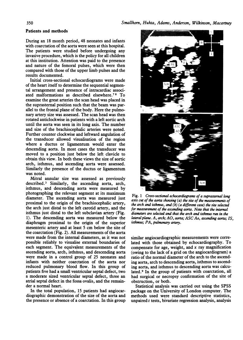
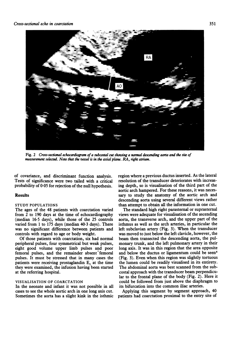
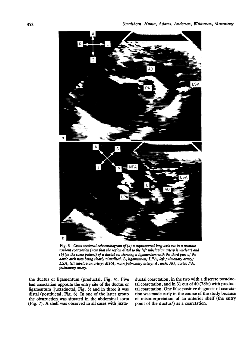
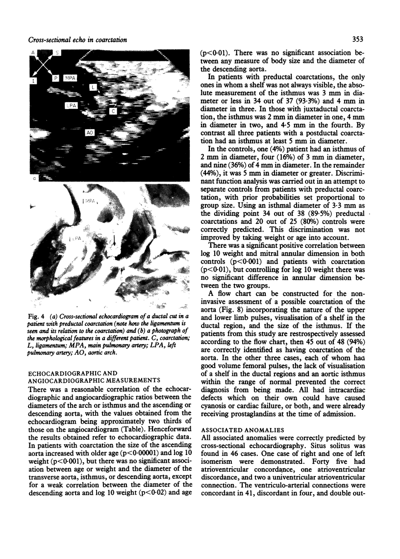
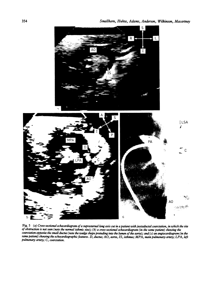
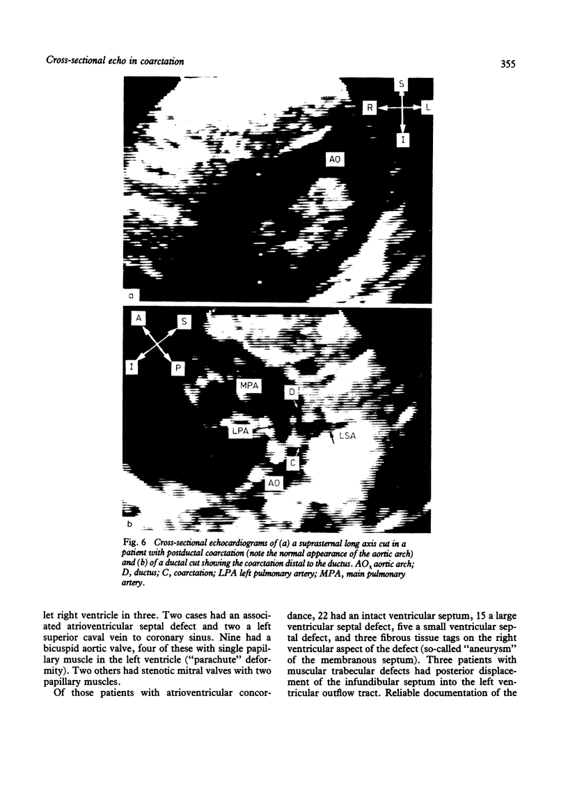
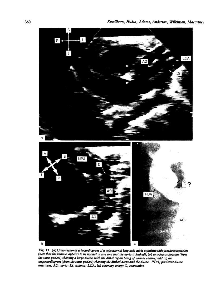
Images in this article
Selected References
These references are in PubMed. This may not be the complete list of references from this article.
- Becker A. E., Becker M. J., Edwards J. E. Anomalies associated with coarctation of aorta: particular reference to infancy. Circulation. 1970 Jun;41(6):1067–1075. doi: 10.1161/01.cir.41.6.1067. [DOI] [PubMed] [Google Scholar]
- Deverall P. B., Taylor J. F., Sturrock G. S., Aberdeen E. Coarctation-like physiology with cerebral arteriovenous fistula. Pediatrics. 1969 Dec;44(6):1024–1028. [PubMed] [Google Scholar]
- Ho S. Y., Anderson R. H. Coarctation, tubular hypoplasia, and the ductus arteriosus. Histological study of 35 specimens. Br Heart J. 1979 Mar;41(3):268–274. doi: 10.1136/hrt.41.3.268. [DOI] [PMC free article] [PubMed] [Google Scholar]
- Huhta J. C., Smallhorn J. F., Macartney F. J. Two dimensional echocardiographic diagnosis of situs. Br Heart J. 1982 Aug;48(2):97–108. doi: 10.1136/hrt.48.2.97. [DOI] [PMC free article] [PubMed] [Google Scholar]
- Leanage R., Taylor J. F., de Leval M. R., Stark J., Macartney F. J. Surgical management of coarctation of aorta with ventricular septal defect. Multivariate analysis. Br Heart J. 1981 Sep;46(3):269–277. doi: 10.1136/hrt.46.3.269. [DOI] [PMC free article] [PubMed] [Google Scholar]
- Marquis R. M., Godman M. J. Nomenclature of the ductus arteriosus. Br Heart J. 1983 Mar;49(3):288–288. doi: 10.1136/hrt.49.3.288. [DOI] [PMC free article] [PubMed] [Google Scholar]
- Rudolph A. M., Heymann M. A., Spitznas U. Hemodynamic considerations in the development of narrowing of the aorta. Am J Cardiol. 1972 Oct;30(5):514–525. doi: 10.1016/0002-9149(72)90042-2. [DOI] [PubMed] [Google Scholar]
- Sahn D. J., Allen H. D., McDonald G., Goldberg S. J. Real-time cross-sectional echocardiographic diagnosis of coarctation of the aorta: a prospective study of echocardiographic-angiographic correlations. Circulation. 1977 Nov;56(5):762–769. doi: 10.1161/01.cir.56.5.762. [DOI] [PubMed] [Google Scholar]
- Sinha S. N., Kardatzke M. L., Cole R. B., Muster A. J., Wessel H. U., Paul M. H. Coarctation of the aorta in infancy. Circulation. 1969 Sep;40(3):385–398. doi: 10.1161/01.cir.40.3.385. [DOI] [PubMed] [Google Scholar]
- Smallhorn J. F., Anderson R. H., Macartney F. J. Cross-sectional echocardiographic recognition of interruption of aortic arch between left carotid and subclavian arteries. Br Heart J. 1982 Sep;48(3):229–235. doi: 10.1136/hrt.48.3.229. [DOI] [PMC free article] [PubMed] [Google Scholar]
- Smallhorn J. F., Anderson R. H., Macartney F. J. Two dimensional echocardiographic assessment of communications between ascending aorta and pulmonary trunk or individual pulmonary arteries. Br Heart J. 1982 Jun;47(6):563–572. doi: 10.1136/hrt.47.6.563. [DOI] [PMC free article] [PubMed] [Google Scholar]
- Smallhorn J. F., Huhta J. C., Anderson R. H., Macartney F. J. Suprasternal cross-sectional echocardiography in assessment of patient ducts arteriosus. Br Heart J. 1982 Oct;48(4):321–330. doi: 10.1136/hrt.48.4.321. [DOI] [PMC free article] [PubMed] [Google Scholar]
- Smallhorn J., Tommasini G., Deanfield J., Douglas J., Gibson D., Macartney F. Congenital mitral stenosis. Anatomical and functional assessment by echocardiography. Br Heart J. 1981 May;45(5):527–534. doi: 10.1136/hrt.45.5.527. [DOI] [PMC free article] [PubMed] [Google Scholar]
- Sutherland G. R., Godman M. J., Smallhorn J. F., Guiterras P., Anderson R. H., Hunter S. Ventricular septal defects. Two dimensional echocardiographic and morphological correlations. Br Heart J. 1982 Apr;47(4):316–328. doi: 10.1136/hrt.47.4.316. [DOI] [PMC free article] [PubMed] [Google Scholar]


