Figure 3.

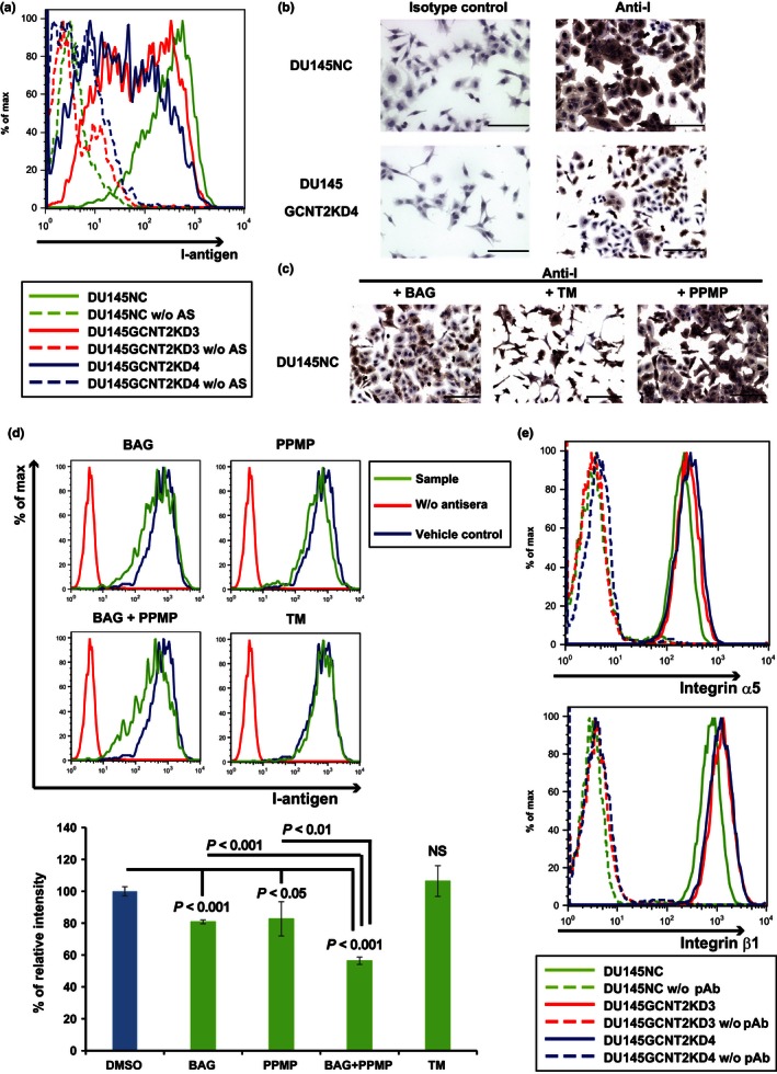
I‐branching N‐acetylglucosaminyltransferase (GCNT2) expression regulates I‐antigen presentation on prostate cancer cells. Cell surface I‐antigen expression was determined using flow cytometry (FC) and immunocytochemistry with anti‐I antigen human antisera (AS). (a) DU145GCNT2KD3 and DU145GCNT2KD4 showed decreased cell surface I‐antigen expression in FC analyses. (b) DU145NC and DU145GCNT2KD4 cells were cultured on glass slides and were stained with anti‐I antigen human antisera or the human IgM isotype control. Brown indicates I‐antigen expression; blue indicates nuclear staining. DU145GCNT2KD4 cells had strongly reduced I‐antigen expression. DU145 cells were cultured with benzyl‐α‐GalNAc (BAG), tunycamycin (TM), or DL‐threo‐1‐Phenyl‐2‐palmitoylamino‐3‐morpholino‐1‐propanol hydrochloride (PPMP) for 48 h. I‐antigen expression was determined using immunocytochemistry (c) and FC (d). I‐antigen expression was significantly reduced in BAG‐treated cells and PPMP‐treated cells. TM‐treated cells had no effect on I‐antigen presentation. Co‐treatment with BAG and PPMP strongly reduced I‐antigen expression compared with either treatment alone. Population comparison was carried out using Flowjo software. Assays were carried out in triplicate. (e) Integrin expression was determined using FC, and expression of α5 and β1 integrins was similar in DU145NC and GCNT2 knockdown cell lines. NS, not significant; pAb, polyclonal antibody. Scale bar = 200 μm.
