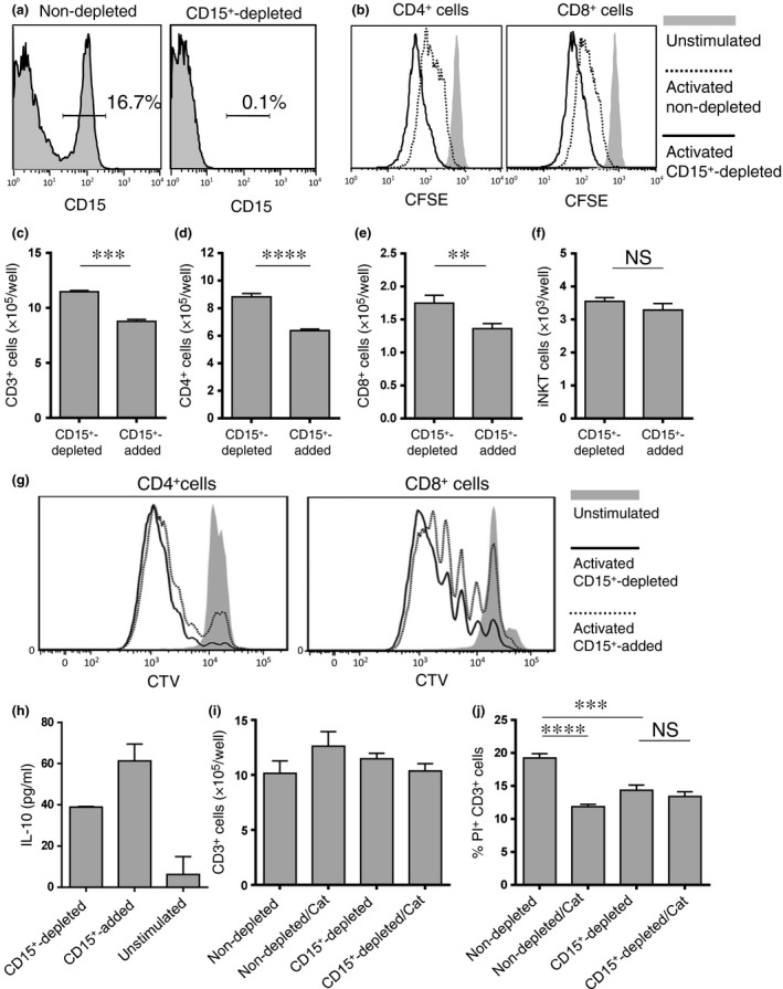
Figure 5.

Granulocytic‐myeloid‐derived suppressor cells (G‐MDSC) suppress autologous T cell proliferation. (a) The percentage of CD15+ cells in HLA‐DR− Lin− fraction of whole peripheral blood cells (PBC) from a head and neck squamous cell carcinoma (HNSCC) patient before (left) and after (right) depletion by CD15 magnetic beads. (b) PBC depleted or not depleted of CD15+ cells were stained with carboxy‐fluorescein diacetate succinimidyl ester (CFSE) and stimulated with anti‐CD3/anti‐CD28 antibodies for 48 h. The difference in CD4+ and CD8+ T cell proliferation was assessed by CFSE dilution. PBC depleted of or with added CD15+ cells were stained with CellTrace Violet and stimulated with anti‐CD3/anti‐CD28 antibodies for 72 h. (c–e) The differences in CD3+, CD4+ and CD8+ cell number were counted, and (g) CD4+ and CD8+ cell proliferation was assessed by CTV dilution. (h) Levels of interleukin (IL)‐10 were measured in supernatants of T cell cultures at 24 h. (f) PBC depleted of or with added CD15+ cells were cultured for 5 days stimulated with αGalCer, and invariant NKT (iNKT) cell number was counted. (i, j) PBC depleted or not depleted CD15+ cells were stimulated with anti‐CD3/anti‐CD28 antibodies with or without catalase (Cat) for 72 h. The difference in CD3+ cell number was counted and cell death was assessed by the percentage of PI+ cells in CD3+cells. **P < 0.01, ***P < 0.001, ****P < 0.0001.
