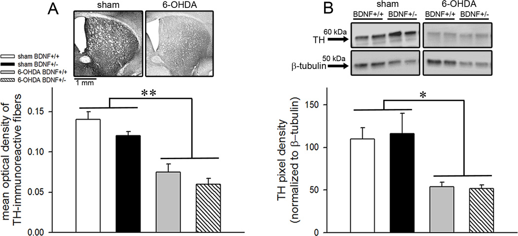
Figure 2.
Tyrosine hydroxylase (TH) protein expression in the dorsal striatum. (A) Representative sections from the dorsal striatum show TH immunohistochemical staining from BDNF+/+ mice either infused with vehicle or 6-OHDA (top). Bar charts depicting optical density values show reduced TH-immunoreactivity in both BDNF+/+ and BDNF+/− mice (bottom). (B) Immunoblots depicting striatal TH expression from duplicate samples representative of all measured bands (top). TH-immunoreactive band (60 kDa) was detected with a monoclonal anti-TH antibody. Blot densities normalized to β-tubulin (gel-loading control) show ~53% decline in TH protein expression in both genotypes infused with 6-OHDA indicating partial dopamine depletion (bottom). TH expression did not differ by genotype. All data are Mean±SEM. (*, ** p<0.05, 0.001 main effect of 6-OHDA manipulation).

