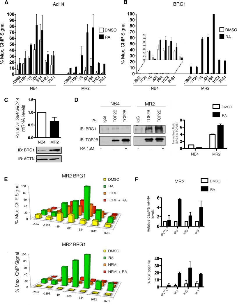
Figure 5. Targeting BRG1 restores sensitivity to RA-induced gene transcription and differentiation.
(A) High-density ChIP tiling of acetylated H4 (AcH4) at the CEBP locus before and after RA treatment (1h) of NB4 and MR2 cells. (B) High-density ChIP tiling of BRG1 at the CEBPB locus before and after RA treatment (1h) of NB4 and MR2 cells. (C) Immunoblot analysis demonstrating differential basal BRG1 protein expression in NB4 and MR2 cells (bottom) with no corresponding increase in BRG1 mRNA levels (top). SMARCA4 is the gene that encodes for the protein known as BRG1. (D) Endogenous co-immunoprecipitation with TOP2B antibody followed by immunoblotting for BRG1 and TOP2B indicates an interaction between BRG1 and TOP2B solely in MR2 cells (left panel). The densitometry values of immunoprecipitated BRG1 were normalized to the amount of immunoprecipitated TOP2B and reported as fold induction over DMSO treated NB4 cells. Densitometry analyses were performed with ImageJ software (right panel). (E) High-density ChIP tiling of BRG1 at CEBPB after treatment with DMSO, RA (1h), the TOP2B inhibitor ICRF (overnight), or a combined treatment of ICRF (overnight pretreatment) and RA (1h) in MR2 cells (top). High-density ChIP tiling of BRG1 at the CEBPB locus after treatment with DMSO, RA (1h), NPM inhibitor (NPMi, overnight), or a combined treatment of NPMi (overnight pretreatment) and RA (1h) in MR2 cells (bottom). (F) Q-RT-PCR analysis of CEBPB mRNA induction in BRG1 MR2-knockdown clones following 8h RA treatment expressed as fold induction over untreated cells after normalization to 18S rRNA levels. Error bars represent the standard error (top) Results of NBT reduction assay performed on BRG1 knockdown MR2 cells treated with RA for 5 days, with retreatment at day 3 (bottom).

