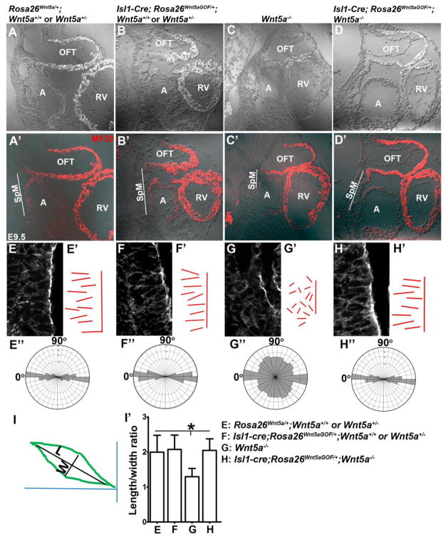
Figure 2.
Rescuing the cell polarity and SpM shortening defects in Wnt5a−/− mutants by Wnt5a over-expression (A′–D′) Immunofluorescent staining for myocardial marker MF20 was performed on sagittally sectioned E9.5 embryos to identify the atrium (A) and outflow tract (OFT). Fluorescent image was overlaid on DIC (differential interference contrast) image shown in (A)–(D). Compared to control embryos (A′), the length of the SpM between the OFT and atrium (outlined by white lines) was significantly shortened in Wnt5a−/− mutants (C′). This defect was rescued by over-expression of Wnt5a throughout the SpM in Isl1-Cre; Rosa26Wnt5aGOF/+; Wnt5a−/− embryos (D′). (E–H′) Phalloidin staining of the sagittal sections also revealed that SHF cells in the caudal SpM of control embryos were elongated and polarized, with their long axes oriented along the dorsal-ventral (D–V) axis of the embryo and perpendicular to the plane of the SpM (E, E′). These cells also display distinct actin filaments largely aligned along the D–V axis (E). In Wnt5a−/− embryos, the SHF cells in the caudal SpM display diminished and disorganized actin filaments, less elongated cell shape, and more randomized orientation of their long axis (G, G′). Isl1-Cre; Rosa26Wnt5aGOF/+ induced homogeneous Wnt5a expression in the SHF significantly rescued the elongation, orientation, and actin polymerization defects in the caudal SHF cells in Wnt5a−/− mutants (H, H′), but had no observable effect on the SHF cells in Wnt5a+/− or Wnt5a+/+ embryos (F, F′). To quantify cell polarity, lines were drawn along the long axis of each cell and represented in E′-H′. The length to width ratios (LWR) and angularity of each SHF cell in the SpM were calculated as depicted in (I). (E″–H″) Analyses with rose.net software indicated that in Rosa26Wnt5a/+ (E″), Isl1-Cre; Rosa26Wnt5aGOF/+; Wnt5a+/− or Wnt5a+/+ (F″) and Isl1-Cre; Rosa26Wnt5aGOF/+; Wnt5a−/− (H″) embryos, majority of the SHF cells (80%, 81% and 79% respectively) in the caudal SpM aligned their long axis within a ±20° arc perpendicular to the plane of the SpM (90°) and along the D–V axis of the embryo (0°). In Wnt5a−/− mutants, however, their orientations were randomized (G″). (I′) Measurement of the LWR of SHF cells in the caudal SpM. Rosa26Wnt5a/+: 1.99±0.48; Isl1-Cre; Rosa26Wnt5aGOF/+; Wnt5a+/− or Wnt5a+/+: 2.07±0.41; Wnt5a−/−: 1.29±0.23; Isl1cre; Rosa26Wnt5aGOF/+; Wnt5a−/−: 2.04±0.34. * indicated statistical significance with p<0.001.

