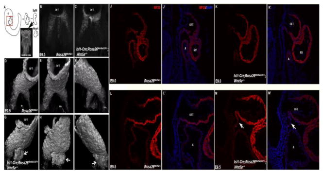
Figure 4.
SHF cells form aberrant bulge in the rostral SpM of Isl1-Cre; Rosa26Wnt5aGOF/+ mutants. (A) Schematic diagram showing how the SpM and distal OFT were isolated for staining and imaging to analyze myocardial differentiation. (B, C) The SpM and OFT from E9.5 control and Isl1-Cre; Rosa26Wnt5aGOF/+ mutant embryos were stained with MF20 and imaged ventrally under confocal microscope. (D–F) In control embryos, 3-D reconstruction of the z-stack images revealed that MF20-positive SHF progenitors were present as two narrow, lateral ridges in the rostral SpM, behind the OFT. (G–I) In Isl1-Cre; Rosa26Wnt5aGOF/+ mutants, however, the MF20 positive cells formed large, abnormal bulges under the OFT (arrows). (J–M′) Sagittal sections of E9.0 (J–K′) and E9.5 (L–M′) control (J & J′; L & L′) and Isl1-Cre; Rosa26Wnt5aGOF/+ mutant (K & K′; M & M′) embryos were stained with MF20 (red) and counterstained with DAPI (blue in J′, K′, L′, M′). The aberrant bulge formation could be observed in the rostral SpM of Isl1-Cre; Rosa26Wnt5aGOF/+ mutants at E9.5 (arrows in M & M′), but not at E9.0.

