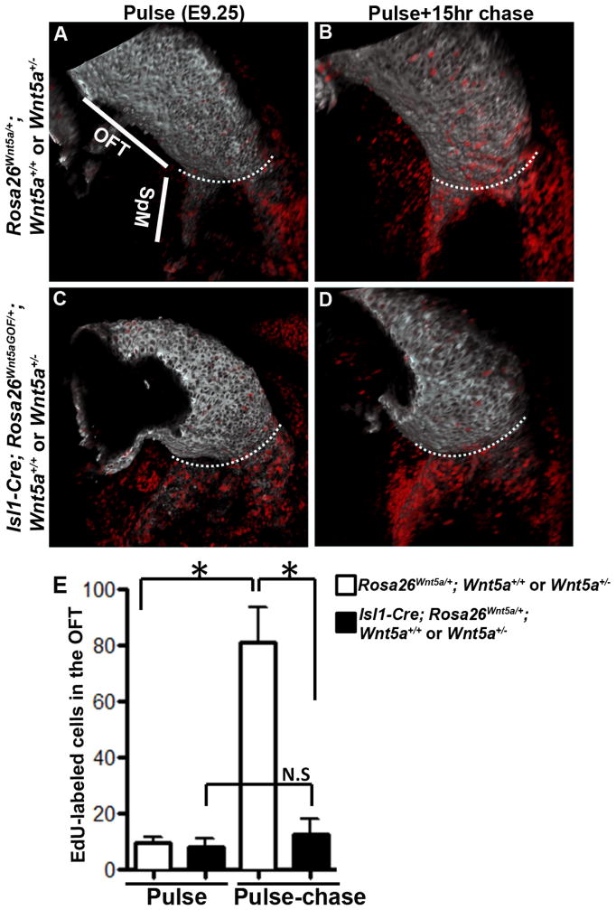
Figure 5.
Deployment of SHF progenitors from the SpM to the OFT is inhibited in Wnt5a GOF mutants. (A, C) E9.25 embryos were pulsed labeled with EdU for 2 hours and processed for whole-mount EdU detection (red) and MF20 staining (grey). Confocal imaging and 3-D reconstruction showed that in both control (A) and Isl1-Cre; Rosa26Wnt5aGOF/+ mutant littermates (C), EdU labeled cells were present almost exclusively in the SpM, but not the OFT. (B, D) EdU pulse labeled E9.25 embryos were chased for 15 hours. After the chase, EdU-labeled cells could be detected in the distal OFT in control (B) but not in Isl1-Cre; Rosa26Wnt5aGOF/+ mutant littermates (D). For each image, the white dotted line marks the boundary between the OFT and the SpM. (E) For quantitative analysis of the total number of EdU-labeled cells in inferior OFT myocardium, 4–7 embryos from each group were imaged and EdU-positive cells were counted and analyzed pairwise with student t test.

