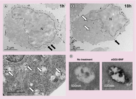
Figure 4. . Iron oxide nanoparticles are internalized after anti-GD2-Bionized NanoFerrite treatment of neuroblastoma cells.
Transmission electron microscope (TEM) images of CHLA-20 cells shows membrane-bound nanoconstructs ([A], black arrows) after 1-h incubation with anti-GD2-BNF, and intracytoplasmic nanoconstructs ([B], white arrows) after 18 h. (C) OsO4 counterstained TEM shows intravesicular electron dense nanoparticles (white arrows) in CHLA-20 after 18 h. (D) Iron staining TEM of intravesicular contents after 18-h culture of CHLA-20 cells treated or not with anti-GD2-BNF. Representative images of two experimental repeats.
BNF: Bionized NanoFerrite; N: Nucleus.

