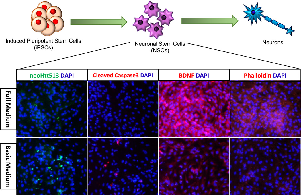
Figure 2. Diagram of iPSC differentiation and potential endpoints at the NSC stage.
Human HD iPSCs were differentiated into NSCs, then neurons and glia. At the NSC stage, we observed disease-associated phenotypes. When stressed (change from full medium to basic medium without growth factors) NSCs derived from HD iPSCs demonstrated upregulation of the N-terminal 513aa Htt fragment (recognized by neoHtt513 antibody) and cleaved caspase-3 and downregulation of BDNF and phalloidin labelled F-actin.

