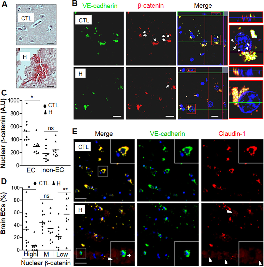
Figure 7. Defective Claudin-1 expression in brain microvascular ECs of hemorrhagic stroke patients.
(A) H&E staining demonstrating petechial hemorrhages in brain sections of these patients. H, hemorrhagic stroke patients; CTL, controls. Scale bars, 30µm. (B) Representative micrographs of co-immunostaining of VE-cadherin (green) and β-catenin (red) of brain sections by confocal microscopy. Nuclei were counterstained with DAPI (blue). Arrows, nuclear expression of β-catenin in microvascular ECs. The merged image displayed the ortho view of Z-stack. The typical cells in red rectangle were blown-out and shown in XY and XZ planes. Scale bars, 10µm. (C, D) Quantification of nuclear β-catenin expression in brain microvascular ECs. The fluorescent intensity of β-catenin in nuclei was quantified using the software Zen2 from Zeiss.. Nuclear β-catenin expression in ECs but not in non-ECs of hemorrhagic tissues was markedly decreased compared to control tissues (C). **, P = 0.0037 (Mann-Whitney 2-tailed test). ns, not significant. Based on the median value of fluorescent intensity of nuclear β-catenin in ECs of control samples, three levels, High [> (median value+1/2SD)], Medium (M), and Low [< (median value-1/2SD)] were set. High level of nuclear β-catenin expression was observed in ~35% of brain ECs in control tissues whereas < 7% in hemorrhagic samples (D). ~60% brain ECs in hemorrhagic patient samples expressed only minimal (Low) nuclear β-catenin (D). *, P = 0.014; **, P = 0.0076 (Mann-Whitney 2-tailed test). ns, not significant. Bars represent mean. (E) Representative micrographs of co-immunostaining of VE-cadherin (green) and Claudin-1 (red) of brain sections from controls, and hemorrhagic stroke patients. Arrowheads, red blood cells (light red), indicating hemorrhages; Arrow, microvessel with absent Claudin-1 expression. Please note the leaked red blood cells (arrowhead) surrounding the microvessel absent Claudin-1 expression shown in the insert, indicating BBB breakdown. Scale bars, 30µm.

