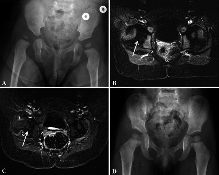
Fig. 5A–D.
The following is a case example of a pMRI-altered treatment course. (A) A 9-month-old girl had right hip DDH as demonstrated on AP pelvis radiograph. She subsequently underwent arthrogram, closed reduction, and spica casting. (B) The immediate postoperative pMRI demonstrated globally decreased perfusion of her right femoral epiphysis (arrow). Her cast was immediately removed and she had a repeat arthrogram and closed reduction 1 week later, at which time she was casted into less abduction. (C) Postreduction pMRI following the second attempt demonstrated symmetric enhancement of the vascular canals throughout the entire epiphysis and of the secondary ossification center (arrow). (D) Most recent AP pelvis radiograph demonstrates excellent interval femoral head development without signs of AVN. Of note, this patient is not included in the reported pMRI cohort as she was treated after the study’s timeframe.

