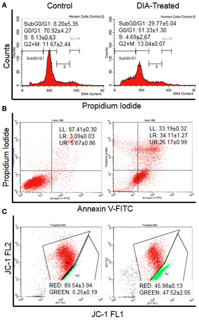
Figure 3.

Flow cytometry analysis of (A) Cell cycle analysis by staining the DNA using propidium iodide, (B) Annexin V analysis for the detection of externalization of phosphatidylserine (LL, viable; LR, Early apoptosis; UR, Late apoptosis), and (C) JC-1 analysis for the detecting the change of mitochondrial membrane potential in HeLa cells (Red: Aggregates; Green: Monomers) after 48 h of treatment with 15 μg/mL of DIA.
