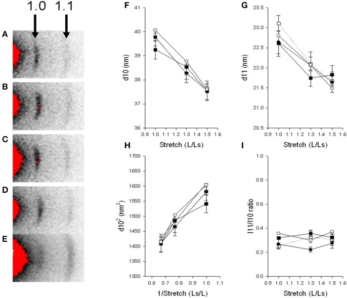
Figure 6.
Small angle X-ray diffraction. Relaxed immotile mutants (D, Filled circles), their control siblings (C, open circles) at 6 dpf, larvae treated with Tricaine between 17 and 24 hpf and recovered until 6 dpf (B, filled squares) and their controls kept in Embryo Medium until 6 dpf (A, open squares). Panels (F,G) shows the spacing (d10, d11) of the 1.0 and 1.1 reflections at different degrees of stretch, (H) shows the relationship between d1.02 and inverted value of stretch. Panel (I) shows the intensity ratio of the 1.1 and 1.0 reflection at different degrees of stretch. n = 4–6 except in the controls kept in Embryo Medium until 6 dpf, where n = 1. Panel (E) shows a relaxed immotile mutant sample in rigor, displaying an increase in the outer 1.1 reflection, the 1.1/1.0 intensity ratio was 3.67 compared to about 0.4 in samples under normal conditions (D,I).

