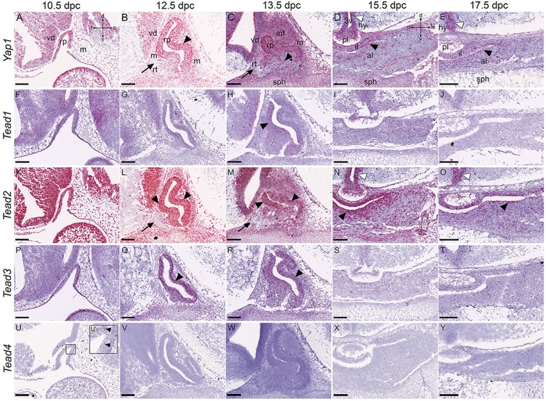
Figure 4.

Expression of the Hippo pathway effectors during embryonic development of the pituitary gland. RNAscope mRNA in situ hybridization using probes against Yap1, Tead1, Tead2, Tead3, and Tead4 on sections of wild type CD1 embryos between 10.5dpc and 17.5dpc. (A–E) Yap1 transcripts are detected in neural tissue, mesenchyme and Rathke's pouch epithelium between 10.5dpc and 13.5dpc (A–C, arrowheads indicating RP expression). Note the dorsal expression bias at 12.5dpc and 13.5dpc and absence of transcripts in the rostral tip (arrows in B,C). Transcripts persist in all tissues at 15.5dpc and 17.5dpc especially in the periluminal region (black arrowhead in D) and epithelium surrounding the third ventricle (white arrowheads in D,E). (F–Y) Expression of Tead1, Tead2, Tead3, and Tead4 encoding TEAD transcription factors. Tead1 and Tead3 transcripts are detectable in all tissues at low levels (F–J,P–T), higher in RP (arrowheads in F,H,P–R). Tead2 is highly expressed in all tissues at 10.5dpc (K), and from 12.5dpc becomes restricted to the ventral diencephalon in neural tissue (L,M) and to the epithelium surrounding the third ventricle (white arrowheads in N,O). Tead2 is strongly expressed in the periluminal epithelium of the anterior pituitary primordium (black arrowheads L–O) but excluded from the rostral tip (arrows L,M). Tead4 transcripts are barely detectable (U–Y). Abbreviations: rp, Rathke's pouch; vd, ventral diencephalon; m, mesenchyme; inf, infundibulum; sph, sphenoid; rt, rostral tip; pl, posterior lobe; al, anterior lobe; il, intermediate lobe; hy, hypothalamus; 3v, third ventricle. Sagittal sections between 10.5dpc and 13.5dpc and frontal between 15.5dpc and 17.5dpc. Axes in (A) applicable to (A–C,F–H,K–M,P–R,U–W: d, dorsal; v, ventral; r, rostral; c, caudal). Axes in (D) applicable to (D,E,I,J,N,O,S,T,X,Y: d, dorsal; v, ventral; ri, right; le, left). Scale bars 200 μm.
