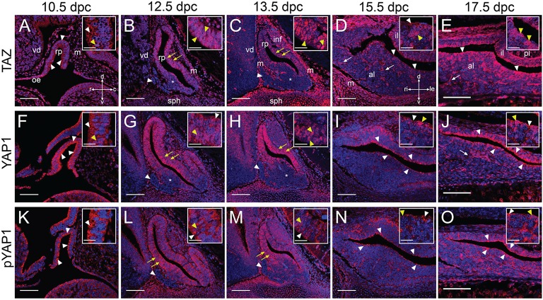
Figure 5.
Expression of YAP1 and TAZ proteins during embryonic development. Immunofluorescence using specific antibodies against total TAZ protein, total YAP1 protein and phosphorylated YAP1 (S127) in red. Nuclei are counterstained with DAPI (blue). (A–J) Localization of effectors TAZ (A–E) and YAP1 (F–J) using antibodies recognizing total protein. Note the nuclear localization in periluminal cells of Rathke's pouch (yellow arrowheads in B,C,G,H, white arrowheads in D,E,I,J). No YAP1/TAZ proteins are detected in the rostral tip (arrowheads in B,C,G,H) and there is a reduction in expression in ventral regions (asterisks in B,C,G,H). Note expression in structures resembling capillaries from 15.5dpc (arrows in D,E,J). (K–O) Immunofluorescence to detect the phosphorylated form of YAP1 at S127. Protein detection indicates Hippo kinase cascade activity at all stages, primarily in periluminal RP epithelium (white arrowheads in K,N,O). Note the ventral bias of protein localization at 12.5dpc and 13.5dpc (yellow arrows in L,M) and complete absence of protein from the rostral tip (white arrowheads in L,M). Boxed inserts show higher magnifications of the epithelium. Examples of cytoplasmic localization are noted by white arrows and examples of nuclear localization by yellow arrows. Abbreviations: rp, Rathke's pouch; vd, ventral diencephalon; m, mesenchyme; oe, oral ectoderm; inf, infundibulum; sph, sphenoid; rt, rostral tip; pl, posterior lobe; al, anterior lobe; il, intermediate lobe. Sagittal sections between 10.5dpc and 13.5dpc and frontal between 15.5dpc and 17.5dpc. Axes in (A) applicable to (A–C,F–H,K–M: d, dorsal; v, ventral; r, rostral; c, caudal). Axes in (D) applicable to (D,E,I,J,N,O: d, dorsal; v, ventral; ri, right; le, left). Scale bars 100 μm and 20 μm in boxed inserts.

