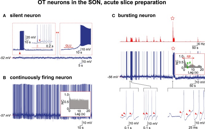
Figure 1.
Electrical activity in OT neurons in hypothalamic acute slices. (A) A silent OT cell displaying rare action potentials (APs, asterisks) was subjected to two depolarizing challenges (insets). Left inset, a brief positive current pulse (50 ms, 0.1 nA; red trace; expanded from the large arrowhead) triggered a 10 s-long decaying burst of APs (the small arrowhead shows the DAP maintaining firing). Right inset, glutamate (GLU) in the perfusion medium (10−5 M, 30 s) induced a strong depolarization triggering a sustained firing. (B) A continuously active OT neuron with irregular firing with no rhythmic drive from the APs autocorrelation analysis (inset). (C) Exceptional example of a truly bursting OT neuron. Top, rate-meter showing the high-frequency burst (HFB) of action potentials (star). Middle, respective raw recording and associated autocorrelogram (inset) showing rhythmic drive. Bottom, expanded traces showing the importance of the EPSPs (arrowheads) in initiating the burst (left) and HFB (right) and triggering the APs within the burst (middle) (note that APs were truncated in the right trace).

