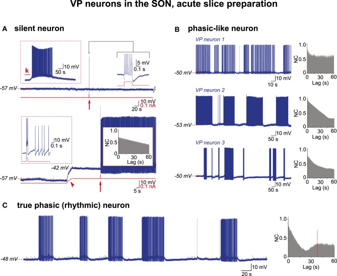
Figure 3.
Electrical activities in VP neurons in hypothalamic acute slices. (A) A silent VP cell displaying action potentials only if subjected to depolarizing challenges. Top trace, left inset, sustained firing in response to kainate (k) in perfusion medium (10−5 M; 30 s); right inset, a train of APs (expanded trace; truncated) in response to a brief positive current pulse (0.2 s, 0.15 nA; red arrow). Bottom trace, the same neuron when depolarized by constant current injection (red trace, arrowhead) now displays a prolonged firing in response to a brief current pulse (0.1 s, 0.1 nA) (left inset, expanded from arrow). Note absence of rhythmic drive (right inset). (B) Three examples of phasic-like activities displayed by VP neurons. Neuron 1, bursts and silent periods of irregular duration. Neuron 2, bursts and silent periods of similar duration. Neuron 3, short and long bursts, irregular silent periods. Autocorrelograms show no rhythmic drive. (C) A truly phasic activity sustained by a rhythmic drive.

