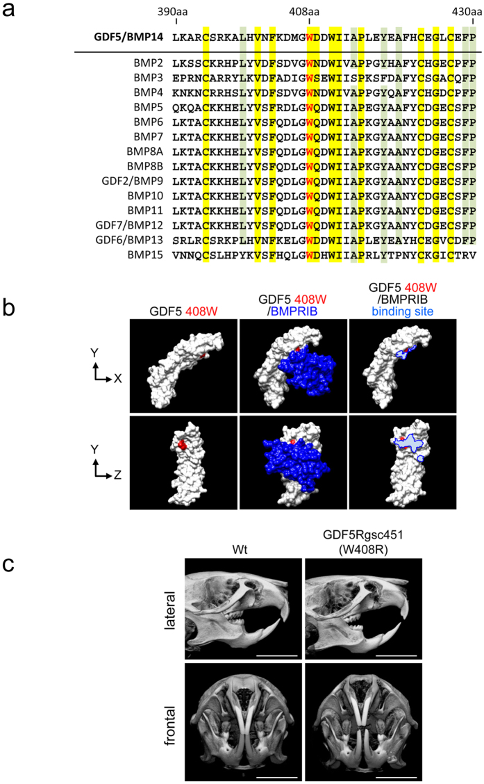
Figure 5. Structural analysis of GDF5 and micro-CT imaging of W408R GDF5 mutant and wild-type mice.
(a) Multiple protein sequence alignment of BMP family. Integrity identity; yellow. High similarity; green. (b) The tertiary structure was assembled using the mouse GDF5 sequence alignment. Amino acid 408 is highlighted in red. (c) Wild-type and GDF5Rgsc451 (W408R) mice were evaluated using a micro-CT scanner. Scare bar: 5,000 μm.

