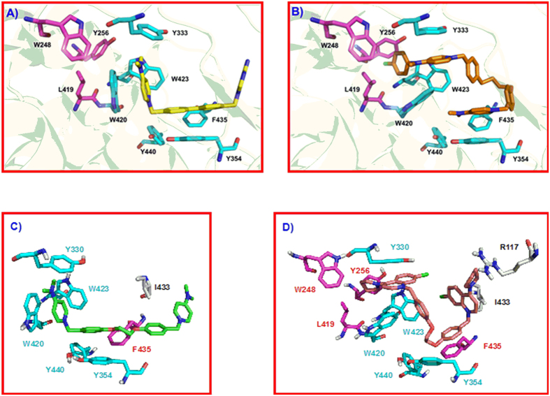
Figure 3.
(A) Crystal structure of ChoKα1/2 complex (PDB ID: 4BR3). Compound 2 (carbon atoms in yellow color) is inserted into the Cho binding site (carbon atoms in cyan color). (B) Crystal structure of ChoKα1/4 complex (PDB ID: 4CG8). Compound 4 (carbon atoms in orange color) is inserted into the Cho binding site (carbon atoms in cyan color) and in an additional binding site (carbon atoms in magenta color) that has been open by a conformational change of Tyr256, Tyr333, and Trp420 sidechains, induced by the insertion of compound 4 into the enzyme. (C) Resulting pose of compound 10a (carbon atoms in green color) in the Cho binding site of the ChoKα1/2 crystal structure, and D) Resulting pose of compound 10l (B, carbon atoms in yellow color) in the ChoK binding site of the ChoKα1/4 crystal structure.

