
Figure 5. LLC produce TNF-α mRNA in the old mouse brain.
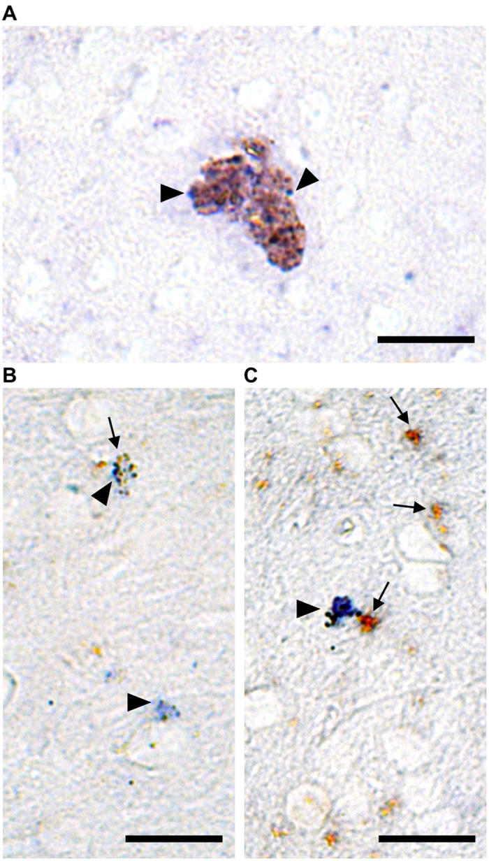
(A–C) Bright field photomicrograph shows brownish PLIN+ LLC expressing TNF-α mRNA (blue) in the cerebral cortex. In situ hybridization for TNF-α mRNA, positively detected using alkaline phosphatase (arrowheads indicate blue staining), was done in combination with immunoperoxidase for PLIN (arrows indicate brown staining) in old brain tissue. Scale bars: 20 μm.