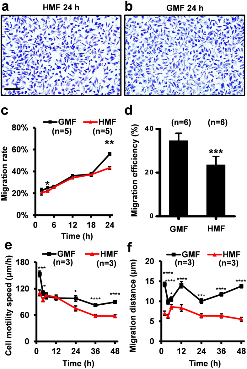
Figure 3. Decreased cell migration in the HMF.
Representative crystal violet staining of the migrated cells on the transwell inserts in the HMF (a) and GMF (b) at 24 h. (c) The migration rate of the HMF-exposed cells was significantly slower than the GMF controls at 4 h and 24 h. (d) The migration efficiency of the cells incubated in the HMF-only environment was significantly less than the controls in the wound-healing assay. (e) The motility speed of the cells exposed to the HMF was significantly slower than the GMF control at 2–4 h and 24–48 h. (f) The migration distance of the HMF-exposed cells was smaller than the GMF control during the 48 h incubation period. Data from separate experiments (n = 5 in c; n = 6 in b; n = 3 in e,f) are shown as mean ± s.e.m. The P values were calculated using a one-way ANOVA. *P < 0.05, **P < 0.01, ***P < 0.001, ****P < 0.0001.

