Figure 1.

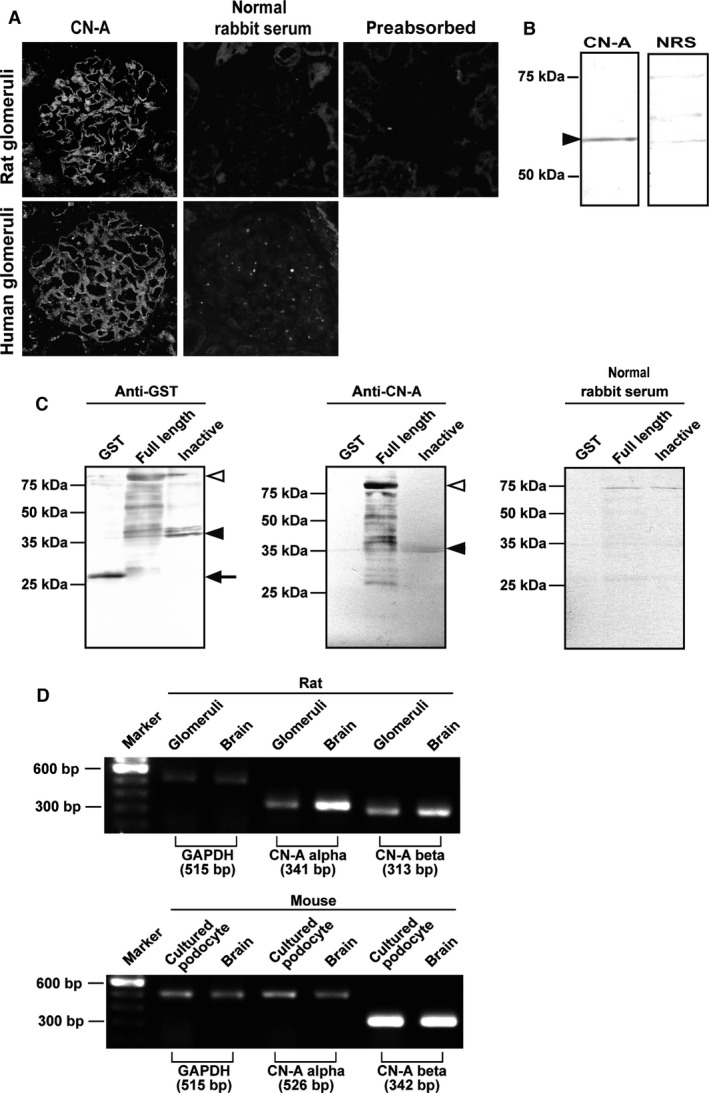
Expression of CN‐A in podocytes in normal glomeruli. (A) Clear positive staining along the capillary wall in the glomeruli of a normal rat kidney section was detected with the rabbit anti‐CN‐A antibody. No specific staining was observed in normal rabbit serum. No positive staining was detected in the antibody preabsorbed with the GST‐fusion protein of full‐length CN‐A, showing that the antibody is specific to CN‐A. Similar staining of CN‐A was detected in the human glomeruli. No staining was observed in the normal rabbit serum in kidney section. (B) A positive band of approximately 60 kDa was detected in a normal rat glomerular lysate with an anti‐CN‐A antibody. No bands were detected with the normal rabbit serum. (C) The antibody recognizes the GST‐fusion protein of full‐length CN‐A. A weak band was detected with the antibody in the GST‐fusion protein of the autoinhibitory domain of CN‐A. (D) mRNA expressions of both CN‐A‐α and CN‐A‐β were detected in normal rat glomeruli (upper panel) and in the murine‐cultured podocyte (lower panel) (brain, cerebrum sample).
