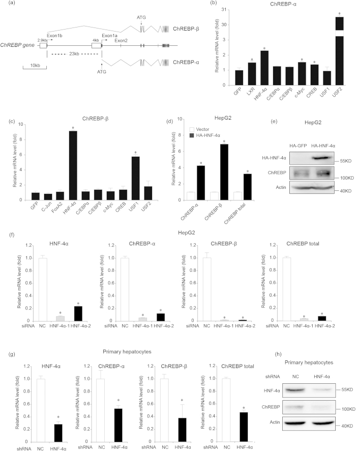
Figure 1. HNF-4α promotes ChREBP-α and ChREBP-β transcription.
(a). Gene structure of human ChREBP-α and ChREBP-β with indication of splice sites and translational start sites (ATG). Note that ChREBP-α and ChREBP-β are transcribed from different promoters separated by 23 kb. (b). Real-time PCR analysis for mRNA levels of ChREBP-α at 48 hours after expression plasmids containing control (GFP) or other cDNAs are transfected in 293T cells. *indicates p < 0.05 when compared with the GFP-transfected sample. (c). Real-time PCR analysis for mRNA levels of ChREBP-β at 48 hours after expression plasmids containing control (GFP) or other cDNAs are transfected in 293T cells. *indicates p < 0.05 when compared with the GFP-transfected sample. (d,e). Real-time PCR analysis for mRNA levels of ChREBP-α, ChREBP-β and total ChREBP (d) and western blot analysis for endogenous ChREBP expression (e) at 48 hours after HA-GFP (vector) or HA-HNF-4α expression plasmids are transfected in HepG2 cells. *indicates p < 0.05 when compared with the vector-transfected sample. Tubulin serves as the loading control. (f).Real-time PCR analysis for mRNA levels of HNF-4α, ChREBP-α, ChREBP-β and total ChREBP at 72 hours after control (NC) or two HNF-4α siRNAs (HNF-4α-1 and HNF-4α-2) are transfected in HepG2 cells. *indicates p < 0.05 when compared with the corresponding NC-transfected sample. (g,h). Real-time PCR analysis for mRNA levels of HNF-4α, ChREBP-α, ChREBP-β and total ChREBP (g) and western blot analysis for endogenous ChREBP expression (h) at 48 hours after control (NC) or HNF-4α shRNAs are transfected in mouse primary hepatocytes. *indicates p < 0.05 when compared with the corresponding NC-transfected sample. Actin serves as the loading control.

